Project Abstract
Objective
Many studies indicate that cargos in exosomes such as microRNAs (miRs) is one of the most promising tools for biomarkers and therapeutic agents because of their stability in the body fluid. Previous studies have shown that airway epithelial cells and lung cancer cells release exosomes with pathological properties when they are exposed to cigarette smoke extract (CSE). However, the functional role of miRs in exosomes released from CSE exposure has never been tested. In this study, we investigated the functional role of exosomal miRs in lung cancer cells in response to CSE exposure.
Methods
A549, human lung cancer cells, were exposed to CSE and three toxic chemicals that are mostly found in CSE (nicotine, acetaldehyde, and benzene) for 24 h. Ultracentrifugation method was used to isolate the exosomes from cell-culture media. The level of miRs in exosomes was measured by quantitative PCR (qPCR). Finally, exosomes from miR23-a over-expressed cells were treated on A549 to measure the cell proliferation and cisplatin chemoresistance.
Results
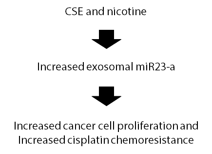
CSE increased the expression level of exosomal miR23-a in A549. Among three selected toxic chemicals, only nicotine increased the expression level of exosomal miR23-a. Interestingly, treatment of exosomes with high-level of miR23-a increased cancer cell proliferation and cisplatin chemoresistance.
Conclusion
Increased expression of exosomal miR23-a by nicotine from CSE exposure promotes the cancer cell proliferation and cisplatin chemoresistance. Therefore, we concluded that exosomal miR23-a is a candidate diagnostic biomarkers of cigarette smoke exposure and potential therapeutic targets for lung cancer. Further study is needed to discover novel function of miR23-a in cancer cells.
Introduction
Smoking cigarette causes 480,000 deaths every year in United States (1 out of every 5 deaths) and causes more than 4 in 5 cases of lung cancer. Non-small cell lung cancer (NSCLC) consists about 85 percentage of lung cancer. It is known that quitting smoking increased 5-year survival rates after the diagnosis of early- and late stage NSCLC to 37% and 34% respectively [1].
Exosomes are cell derived extracellular vesicles that are present in all body fluids. The diameters of exosomes are identified to be ~30-100nm. Exosomes can be both released from the cells and fused with the plasma membrane. Various molecules such as proteins, RNAs, and lipids are packaged in exosomes and can be secreted into diverse body fluids [2]. Therefore, exosomes have great potential as diagnostic tools and biomarkers for various diseases.
MicroRNAs (miRs) are small (19–25 nt), single-stranded, non-coding RNAs that bind to partially complementary sequences to their target messenger RNA (mRNA). miRNAs degrades mRNA and inhibit the translation of target mRNAs [3]. miRNAs are often found in exosomes, which can be taken up by distant cells. Exosomal miRNAs are known to play important role in various disease progression including cancer [4].
Cigarette smoke has been shown to change the expression of genes in airway epithelial cells from smokers [5]. Lung cancer cells exposed to cigarette smoke altered the expression of genes associated with oxidative stress and inflammation [6]. Recent studies indicated that cigarette smoke extract (CSE) induced exosome release in airway epithelial cells. However, the functional role of miRs in exosomes released from CSE exposure has never been tested. In this study we investigated the role of exosomal miR-23a on A549, a model for NSCLC.
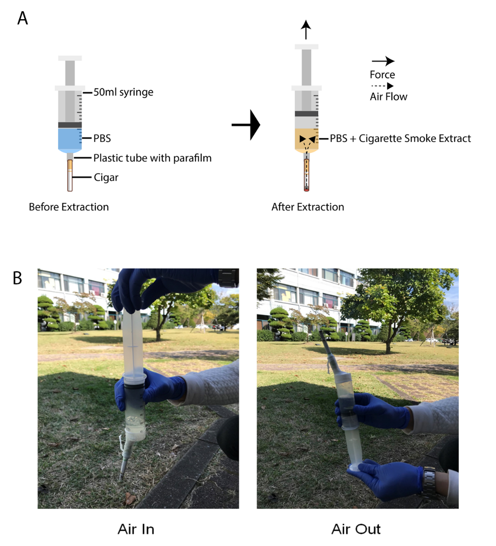
Figure 1. Method for the preparation of CSE. (A) Schematic illustration for extracting cigarette smoke. All smoke passes through cigarette filter and PBS. Soluble chemicals from cigarette smoke remains in PBS (B) Picture for the preparation of CSE by repeating the air pump.
We developed the simple and easy method for extraction of CSE solution. Previous methods for extraction of CSE solution requires vacuum pump and flasks to pull the air through cigarette. Our newly developed method requires 50 ml syringe, phosphate buffered saline (PBS), and parafilm (Figure 1A). When the air flows through the lighted cigarette, the soluble chemicals from cigarette smoke remains in PBS. The air pump was repeated as described in figure 1B.
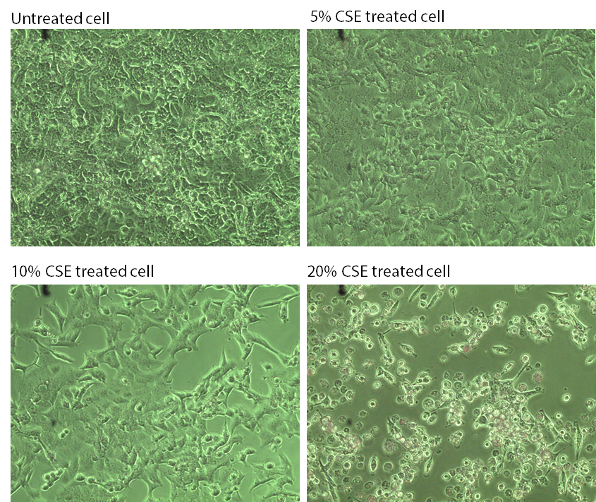
Figure 2. Increased % (V/V) of CSE treatment on A549 changes cell morphology and increases cell death.
In order to find the appropriate working concentration of CSE, we checked the cell condition when % of CSE was increased. 5% CSE had little visible effect on cell condition. 10% CSE changed cell morphology. 20% CSE killed too many cells. Therefore, we selected 10% CSE condition for later experiments.
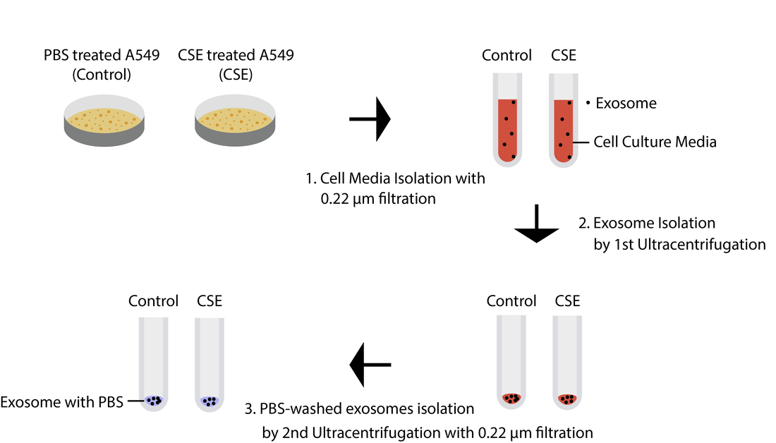
Figure 3. Schematic illustration of exosome isolation from cell culture media by ultracentrifugation. (1) Cell media were isolated from the cell culture (2) Exosomes from cell culture media were isolated by 1st ultracentrifugation (3) PBS was used to wash the exosome by performing 2nd ultracentrifugation and any contamination was filtered by 0.22μm filter
We used ultracentrifugation method to isolate exosome from cell culture media because this method is known to isolate pure exosome (Figure 3). This method do not require adding any chemical solutions which can damage exosomes. Exosomes were successfully isolated from cell culture media. Dr. Lee confirmed the purity of exosomes with exosomal marker by western blot (data not shown).
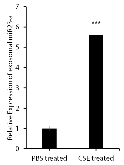
Figure 4. qPCR result shows that exosomal miR-23-a is upregulated by CSE. Triplicate samples were analyzed. (Student T-test from Microsoft excel was used for statistical analysis, *** P ≤ 0.001)
Since cigarette smoking causes lung cancer, we hypothesized that chemicals from cigarette smoke may produce exosomal miRNAs to support cancer cell growth. In order to find the miRNA target we searched many research papers. Among many miRNAs in the exosome, we focused on exosomal miR23-a because miR23-a was recently known for oncogene in pancreatic cancer [7] and lung cancer [8]. Therefore in this study we hypothesized that chemicals from cigarette smoke may affect miR23-a expression. Interestingly, we found that CSE increases expression of exosomal miR23-a more than 5 times that of PBS treated control exosome.
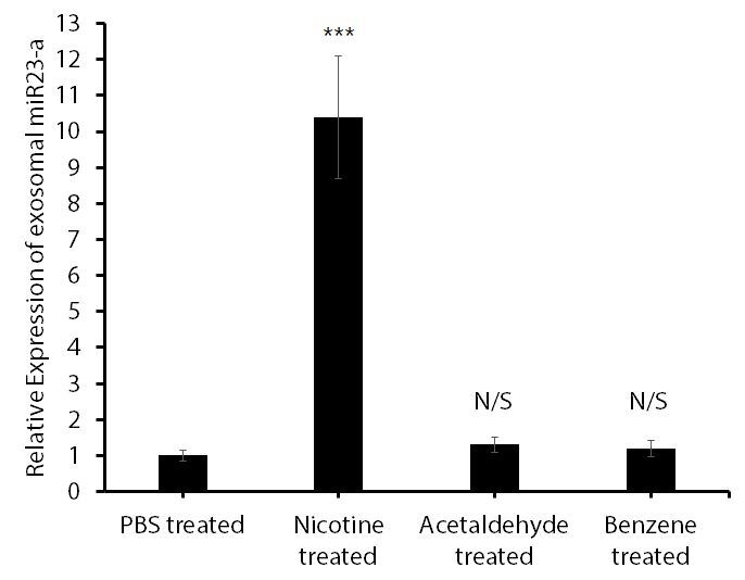
Figure 5. qPCR result shows that exosomal miR23-a is upregulated by nicotine. Nicotine (600 μM), Acetaldehyde (100 μM), Benzene (200 μM) were incubated for 24 h. Triplicate samples were analyzed (Student T-test from Microsoft excel was used for statistical analysis, *** P ≤ 0.001, N/S P > 0.05).
In order to find which chemical among CSE affect miR23-a expression, we separately added the chemicals on A549 cell culture media. Interestingly, only nicotine added cells displayed increase in the expression level of exosomal miR23-a (Figure 5).
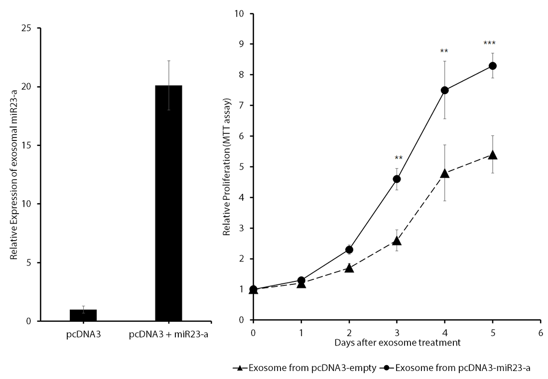
Figure 6. miR23-a overexpression upregulated exosomal miR23-a and increased cell proliferation. Triplicate samples were analyzed (Student T-test from Microsoft excel was used for statistical analysis, ** P≤ 0.01, *** P ≤ 0.001)
Dr. Lee provided the DNA plasmid: pcDNA3 and pcDNA3 + miR23-a. We transfected DNA plasmids on A549 and checked the exosomal miR23-a expression level. Only pcDNA3 + miR23-a plasmid transfected cells produces increased level of exosomal miR23-a (Figure 6 left panel). Next, we tested whether exosomal miR23-a affects cell proliferation. Interestingly, we found that exosome with high level of miR23-a increased cell proliferation (Figure 6 right panel).
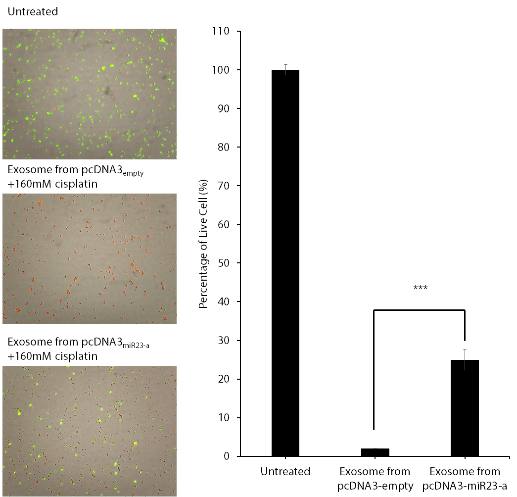
Figure 7. Exosome from miR23-a overexpressed cells increased cisplatin chemoresistance. Cell viability were tested after cisplatin and exosomes were treated for 24 h. Green fluorescent indicates live cells red fluorescent indicates dead cells. Triplicate samples were analyzed (Student T-test from Microsoft excel was used for statistical analysis, *** P ≤ 0.001).
Because exosomal miR23-a increased cell proliferation in our previous experiment, we tested whether exosomal miR23-a affect cisplatin chemoresistance. We tried to test cisplatin because it is commonly used for cancer chemotherapy. Interestingly, exosomal miR23-a increased the number of live cells (Figure 7).
Conclusion and Discussion